Learning Drosophila ventral furrow formation with graph neural networks
Abstract
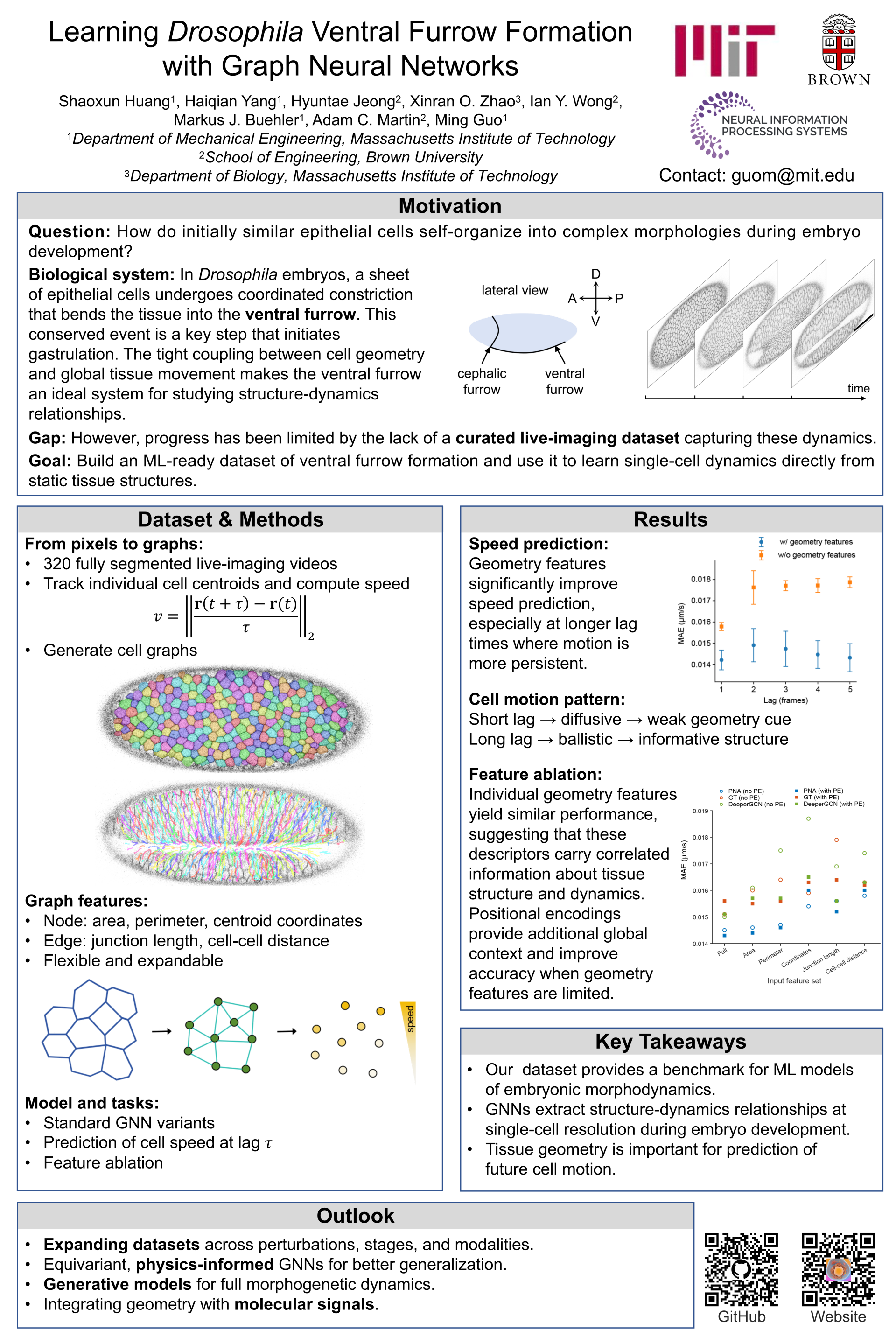
During early embryonic development, a relatively homogeneous population of cells collectively and spontaneously self-organize into complex structure. Decoding the spatiotemporal information of tissue structure and dynamics is a central question in developmental biology. A paradigm process is the Drosophila gastrulation, during which the apical constriction gives rise to the formation of a ventral furrow, a critical step towards forming complex tissue structures. In this process, tissue morphologies and cell dynamics are closely correlated. Many prior biophysical models employ a graph representation of tissue structure, where the cells evolve following the gradient of a pre-defined free energy functional. Here we instead explore using graph neural networks to uncover the relation between tissue structures and dynamics from data, during the dynamical process of Drosophila ventral furrow formation. To do so, we build an experimental video dataset consisting of 320 fully segmented live-imaging movies, capturing the formation of ventral furrow over time. On this dataset, we use a graph neural network to learn individual cell speed from tissue structures, achieving good performance. Our results, along with the dataset, set the stage for future exploration of using graph neural networks for predictive modeling of embryonic development.