Denoising Enhances Visualization of Optical Coherence Tomography Images
Abstract
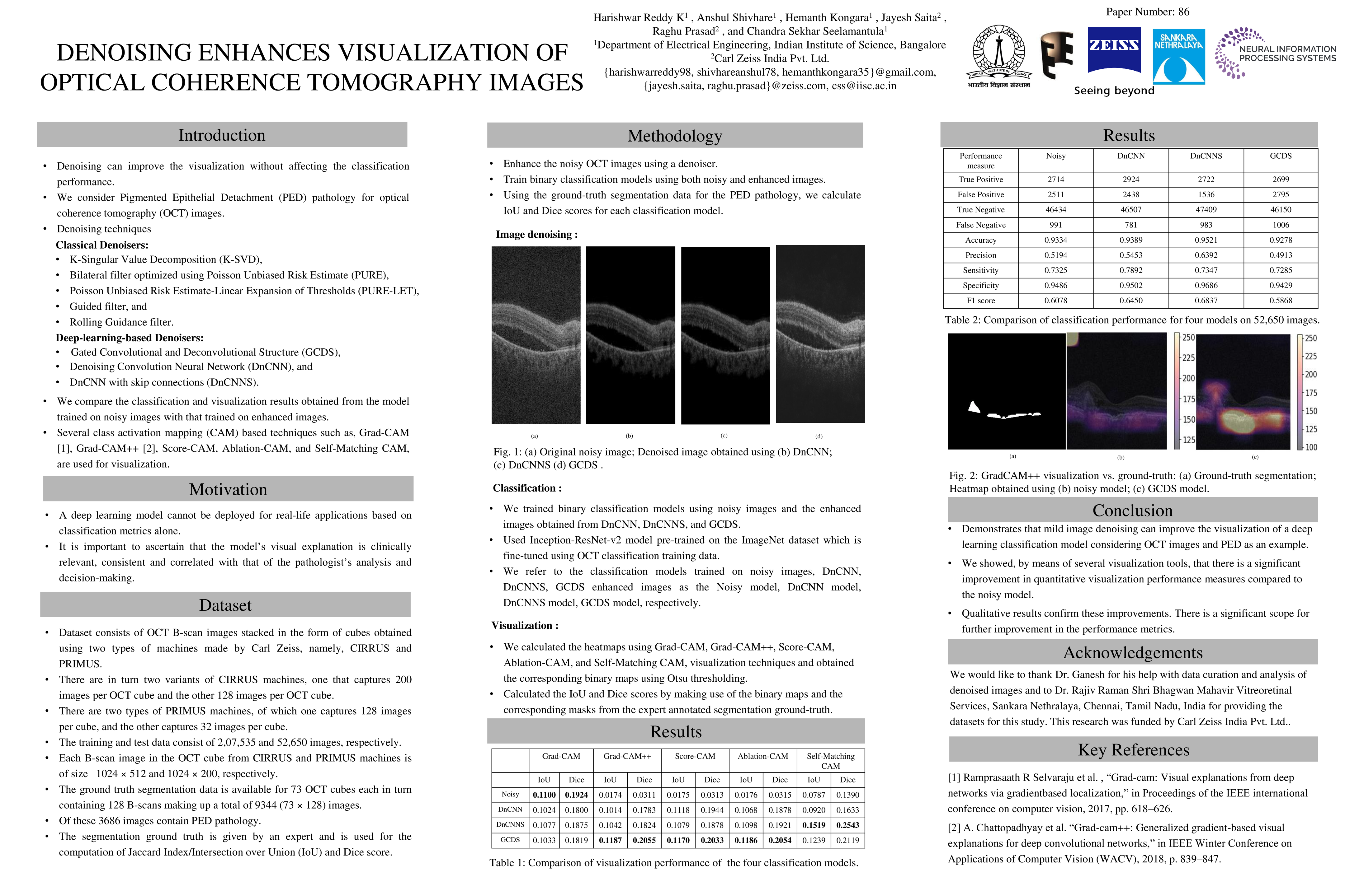
The main aim of this work is to improve the visualization of abnormalities in Optical Coherence Tomography (OCT) images of the human retina. OCT images have substantial noise, which can affect the classification and visualization performance of a neural network. In this work, we show that denoising improves visualization without affecting the classification performance considering the specific pathology of Pigmented Epithelial Detachment (PED). The noise in OCT images may lead to unstable training and poor classification/visualization performance. Hence, the need for image quality enhancement. We consider several image denoising techniques, namely, K-Singular Value Decomposition (K-SVD), Bilateral filter optimized using Poisson Unbiased Risk Estimate (PURE), Poisson Unbiased Risk Estimate - Linear Expansion of Thresholds (PURE-LET), Guided filter, Rolling Guidance filter, Gated Convolutional and Deconvolutional Structure (GCDS), Denoising Convolution Neural Network (DnCNN), and DnCNN with skip connections. We compare the classification and visualization results obtained from the model trained on noisy images with that trained on enhanced images. Several class activation mapping (CAM) based techniques have been developed, for instance, Grad-CAM, Grad-CAM++, Score-CAM, Ablation-CAM, and Self-Matching CAM, which are also the visualization techniques that we employ in this paper. Our results show that denoising improves visualization performance by a factor of approximately10 in Jaccard Index, taking the expert segmentation as the ground-truth.